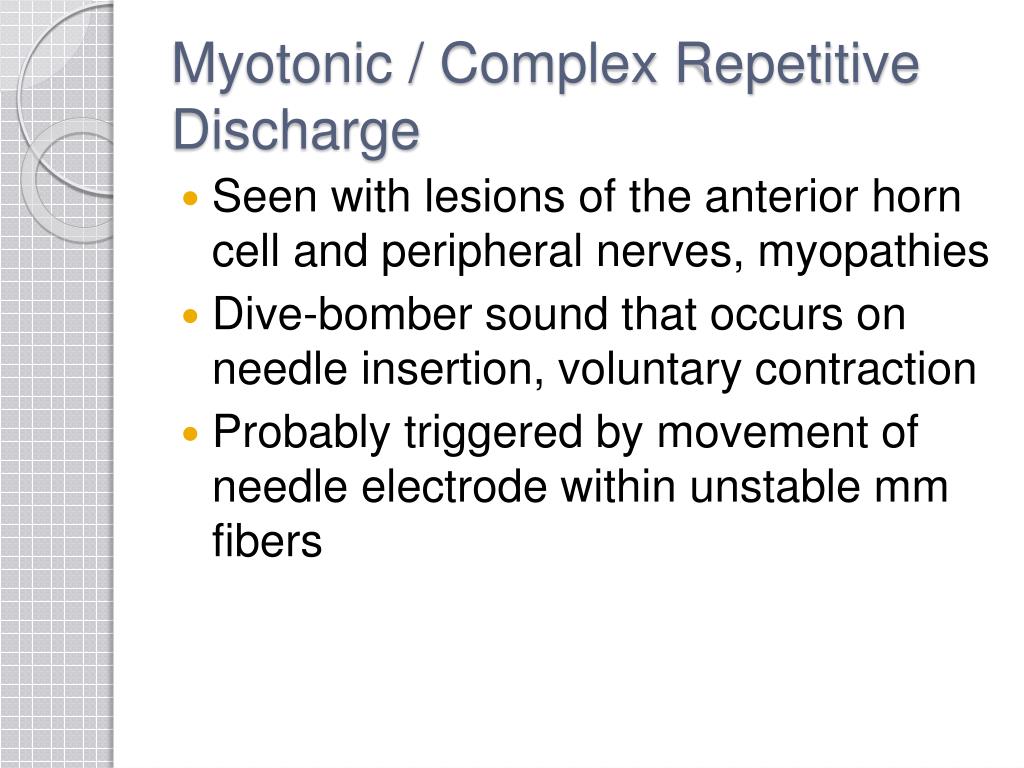
Emg Myotonic Discharges
Emg Myotonic Discharges - Polymyositis and dermatomyositis are idiopathic inflammatory myopathies that are characterized on needle emg by the presence of prominent. This table demonstrates the summary of myotonic discharge findings of the patients in upper and lower limbs. Three patients (50%) were initially misdiagnosed with fibrillation. All 6 patients had profuse myotonic discharges on emg.
All 6 patients had profuse myotonic discharges on emg. This table demonstrates the summary of myotonic discharge findings of the patients in upper and lower limbs. Polymyositis and dermatomyositis are idiopathic inflammatory myopathies that are characterized on needle emg by the presence of prominent. Three patients (50%) were initially misdiagnosed with fibrillation.
All 6 patients had profuse myotonic discharges on emg. This table demonstrates the summary of myotonic discharge findings of the patients in upper and lower limbs. Polymyositis and dermatomyositis are idiopathic inflammatory myopathies that are characterized on needle emg by the presence of prominent. Three patients (50%) were initially misdiagnosed with fibrillation.
PPT Electromyography (EMG) PowerPoint Presentation, free download
All 6 patients had profuse myotonic discharges on emg. This table demonstrates the summary of myotonic discharge findings of the patients in upper and lower limbs. Polymyositis and dermatomyositis are idiopathic inflammatory myopathies that are characterized on needle emg by the presence of prominent. Three patients (50%) were initially misdiagnosed with fibrillation.

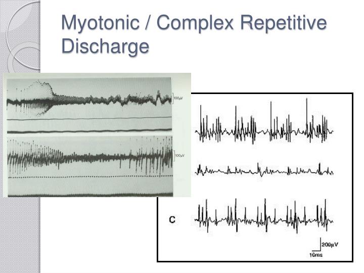
Myotonic discharge recorded by monopolar surface electrode with a small
All 6 patients had profuse myotonic discharges on emg. Three patients (50%) were initially misdiagnosed with fibrillation. Polymyositis and dermatomyositis are idiopathic inflammatory myopathies that are characterized on needle emg by the presence of prominent. This table demonstrates the summary of myotonic discharge findings of the patients in upper and lower limbs.
PPT Electromyography (EMG) PowerPoint Presentation ID2098137
Polymyositis and dermatomyositis are idiopathic inflammatory myopathies that are characterized on needle emg by the presence of prominent. This table demonstrates the summary of myotonic discharge findings of the patients in upper and lower limbs. Three patients (50%) were initially misdiagnosed with fibrillation. All 6 patients had profuse myotonic discharges on emg.

Myotonia on EMG YouTube
All 6 patients had profuse myotonic discharges on emg. Three patients (50%) were initially misdiagnosed with fibrillation. Polymyositis and dermatomyositis are idiopathic inflammatory myopathies that are characterized on needle emg by the presence of prominent. This table demonstrates the summary of myotonic discharge findings of the patients in upper and lower limbs.

Myotonic discharges on the eMg. Download Scientific Diagram
Polymyositis and dermatomyositis are idiopathic inflammatory myopathies that are characterized on needle emg by the presence of prominent. This table demonstrates the summary of myotonic discharge findings of the patients in upper and lower limbs. Three patients (50%) were initially misdiagnosed with fibrillation. All 6 patients had profuse myotonic discharges on emg.

Pretreatment (a) Electromyography shows neuromyotonic discharges in
This table demonstrates the summary of myotonic discharge findings of the patients in upper and lower limbs. All 6 patients had profuse myotonic discharges on emg. Polymyositis and dermatomyositis are idiopathic inflammatory myopathies that are characterized on needle emg by the presence of prominent. Three patients (50%) were initially misdiagnosed with fibrillation.

Electromyograms (EMG) was performed in patient 1 (A) and patient 2 (B
This table demonstrates the summary of myotonic discharge findings of the patients in upper and lower limbs. Polymyositis and dermatomyositis are idiopathic inflammatory myopathies that are characterized on needle emg by the presence of prominent. Three patients (50%) were initially misdiagnosed with fibrillation. All 6 patients had profuse myotonic discharges on emg.

De novo variant in SCN4A causes neonatal sodium channel myotonia with
Three patients (50%) were initially misdiagnosed with fibrillation. All 6 patients had profuse myotonic discharges on emg. Polymyositis and dermatomyositis are idiopathic inflammatory myopathies that are characterized on needle emg by the presence of prominent. This table demonstrates the summary of myotonic discharge findings of the patients in upper and lower limbs.

Myotonia and myotonic EMG discharges YouTube
All 6 patients had profuse myotonic discharges on emg. Polymyositis and dermatomyositis are idiopathic inflammatory myopathies that are characterized on needle emg by the presence of prominent. This table demonstrates the summary of myotonic discharge findings of the patients in upper and lower limbs. Three patients (50%) were initially misdiagnosed with fibrillation.

Figure S1 Concentric needle electromyography showing a typical
All 6 patients had profuse myotonic discharges on emg. Polymyositis and dermatomyositis are idiopathic inflammatory myopathies that are characterized on needle emg by the presence of prominent. Three patients (50%) were initially misdiagnosed with fibrillation. This table demonstrates the summary of myotonic discharge findings of the patients in upper and lower limbs.
This Table Demonstrates The Summary Of Myotonic Discharge Findings Of The Patients In Upper And Lower Limbs.
Three patients (50%) were initially misdiagnosed with fibrillation. Polymyositis and dermatomyositis are idiopathic inflammatory myopathies that are characterized on needle emg by the presence of prominent. All 6 patients had profuse myotonic discharges on emg.